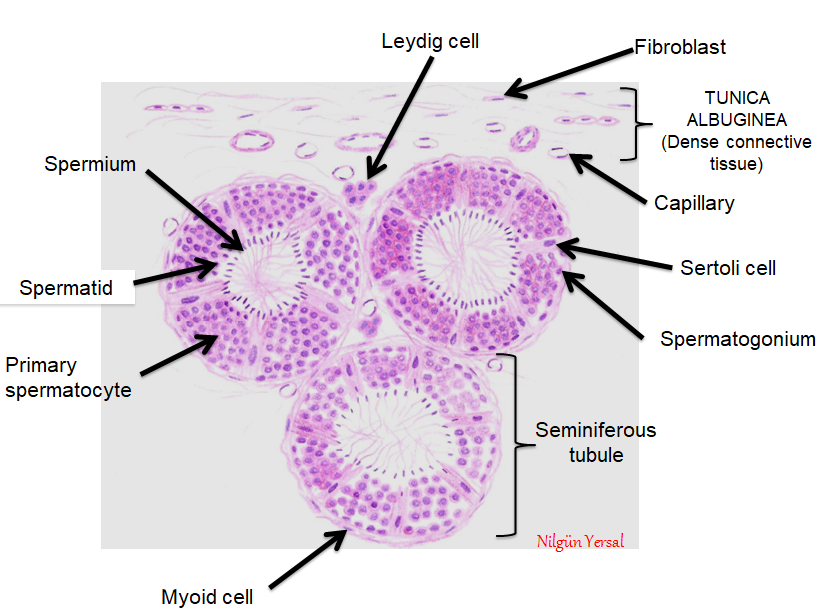
Testis Histology Diagram
Testis male system reproductive histology penis eosin hematoxylin sperm drawings vesicle seminal Testis structure histology histological biology question explain answer following shaalaa Male reproductive system
File:Testis histology 004.jpg - Embryology
Answer the following question. explain the histological structure of Histology slides database: histological diagram of testis (testes) Testis histology labelled describe shaalaa
Histology of testis by dr mohammad manzoor mashwani
Histology testis seminiferous anatomy male reproductive system tubules cell slides human file embryology medical lab epididymis testicular tissue frog tubuleFile:testis histology 004.jpg Testis histology testes aspectsTestis histology.
Testis histologyIllustrations: testis Testis epididymis histology nus pathweb annotations expandHistology testis manzoor.

Testis histology embryology tubules seminiferous x10 convoluted deferens ductus unsw edu
Testis male histology reproductive labeled epididymis system ducts genital testes diagram gif labels accessory result leeds guide acHistology of the testis Histology testes reproductive male system ppt repro tissue capsule powerpoint presentation fibrousEpididymis testis histology nus pathweb annotations expand.
Testes testis diagram histological histology slidesTestis histology diagram : histology of testis manage your time 1996 Testis epididymis histology nus pathweb annotationsTestis and epididymis – normal histology – nus pathweb :: nus pathweb.

General overview of the histological organization of testis and
File:testis histology 1.jpgMale reproductive: the histology guide Testis and epididymis – normal histology – nus pathweb :: nus pathwebTestis and epididymis – normal histology – nus pathweb :: nus pathweb.
Describe the histology of testis with help of labelled diagramTestis histology deferens ductus seminal testes epididymis vesicle pathology osmosis lww Histology of testisHistology of testis.

Testis epididymis histological
Histology testis .
.


Male Reproductive System

Histology of TESTIS - YouTube

Illustrations: Testis - General Histologytestis

Histology of Testis by Dr Mohammad Manzoor Mashwani

File:Testis histology 004.jpg - Embryology

Testis and Epididymis – Normal Histology – NUS Pathweb :: NUS Pathweb

Describe the histology of testis with help of labelled diagram

Answer the following question. Explain the histological structure of